Hot Products
YSX500D 50kW DR system set up and put into service in Cambodia.
YSENMED YSX500D 50kW digital x-ray system has been successfully set up and put into service in a hospital in Cambodia.
YSX056-PE serving as a vehicle-mounted x-ray in the Philippines
YSX056-PE 5.6kW portable x-ray unit has been adapted to fit on a truck, to provide mobile x-ray examination service for remote communities in the Philippines.
X Ray Machine To Zimbabwe
x ray machine, 50KW x ray machine
Microscope To Malawi
Achromatic objectives: 4X、10X、40X(S), 100X(S、Oil) Wide field eyepiece: WF10X(WF16X for option) Eyepiece head: Sliding binocular head inclined at 45° Stage: Double layer mechanical stage size 140X140mm, moving range 75X45mm Focusing: Coaxial coarse and
How do x-ray machines work?
Views : 1096
Author : Yuesen Med
Update time : 2015-11-12 08:00:00
As with many of mankind's monumental discoveries, medical x-ray technology was invented completely by accident.
In 1895, a German physicist named Wilhelm Roentgen made the discovery while experimenting with electron beams in a gas discharge tube. Roentgen noticed that a fluorescent screen in his lab started to glow when the electron beam was turned on.
This response in itself wasn't so surprising -- fluorescent material normally glows in reaction to electromagnetic radiation
but Roentgen's tube was surrounded by heavy black cardboard. Roentgen assumed this would have blocked most of the radiation.
Roentgen placed various objects between the tube and the screen, and the screen still glowed. Finally, he put his hand in front of the tube, and saw the silhouette of his bones projected onto the fluorescent screen. Immediately after discovering x-rays themselves, he had discovered their most beneficial application.
Roentgen's remarkable discovery precipitated one of the most important medical advancements in human history. X ray technology lets doctors see straight through human tissue to examine broken bones, cavities and swallowed objects with extraordinary ease. Modified X ray procedures can be used to examine softer tissue, such as the lungs, blood vessels or the intestines.
How an medical x-ray machine work? An X ray machine is essentially a video camera. Instead of visible light, however,
it uses X rays to expose the film.
it uses X rays to expose the film.
X rays are like light in that they are electromagnetic waves, but they are more energetic so they can penetrate many materials to varying degrees. When the X rays hit the film, they expose it just as light would. Since bone, fat, muscle, tumors and other masses all absorb X rays at different levels, the image on the film lets you see different (distinct) structures inside the body because of the different levels of exposure on the film.
Related News
Read More >>
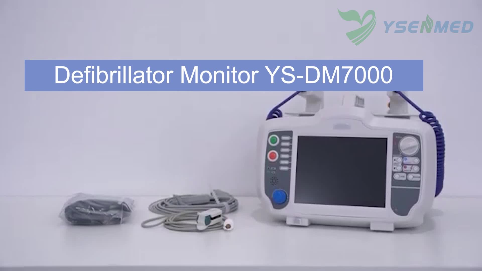
 Introduction video of YSENMED YS-DM7000 Defibrillator Monitor
May .02.2025
Here we share the introduction video of YSENMED YS-DM7000 Defibrillator Monitor.
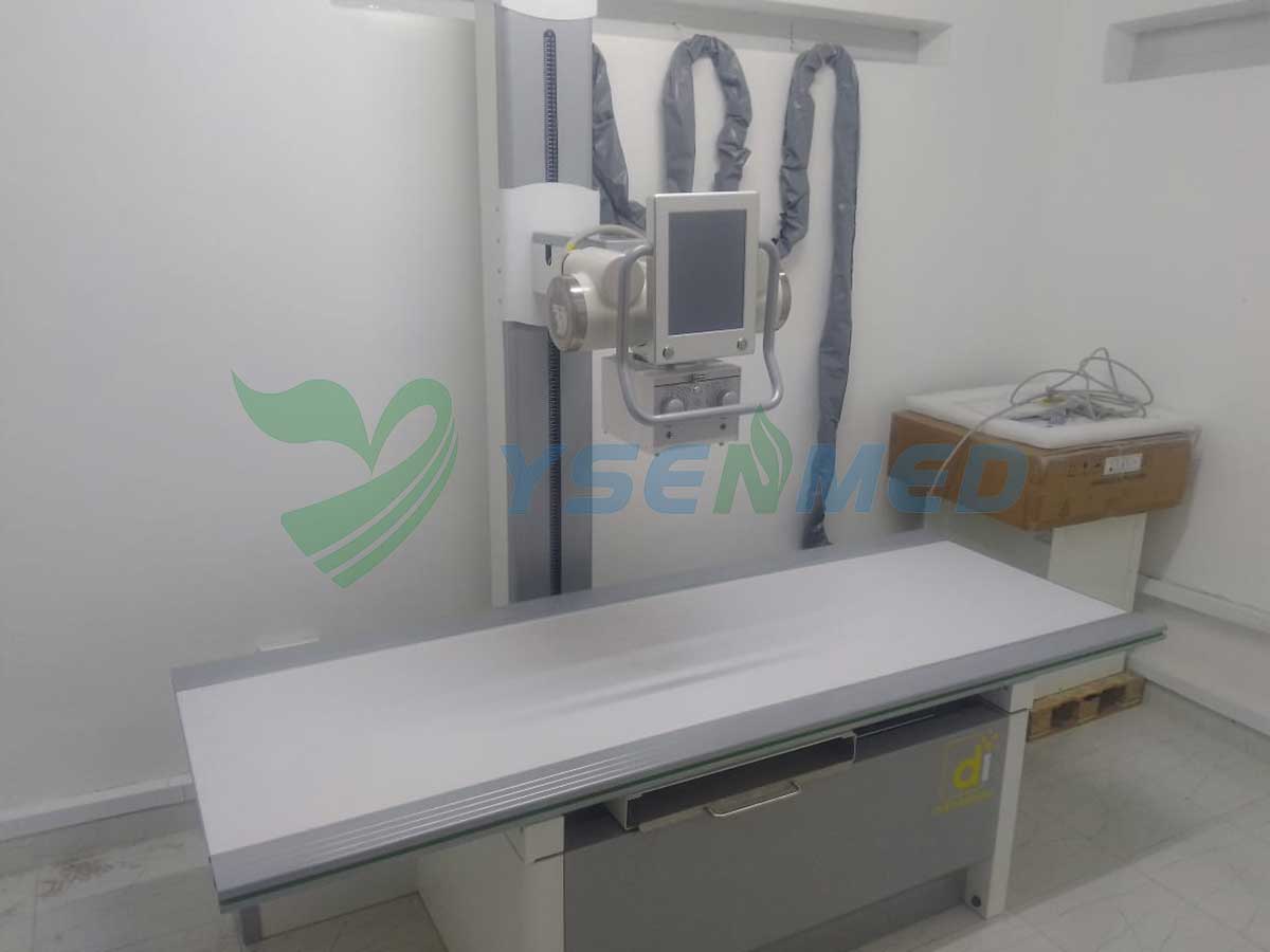
 Kenyan distributor sets up YSX500D DR system and considers re-purchase.
May .01.2025
Our Kenyan distributor has successfully set up the YSX500D 50kW digital x-ray system in a hospital, and considers purchasing another 32kW x-ray system.
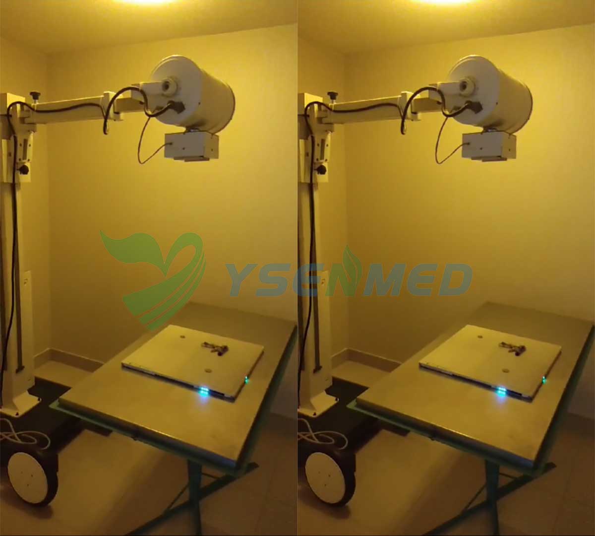
 YSFPD-M1717V VET flat panel detector helps digitize the x-ray in a Peruvian vet clinic
Apr .30.2025
With the professional after-sales assistance from YSENMED team, Peruvian vet has managed to upgrade his mobile analog x-ray system with YSFPD-M1717V VET wilress flat panel detector and the images come out clear and highly satisfactory.
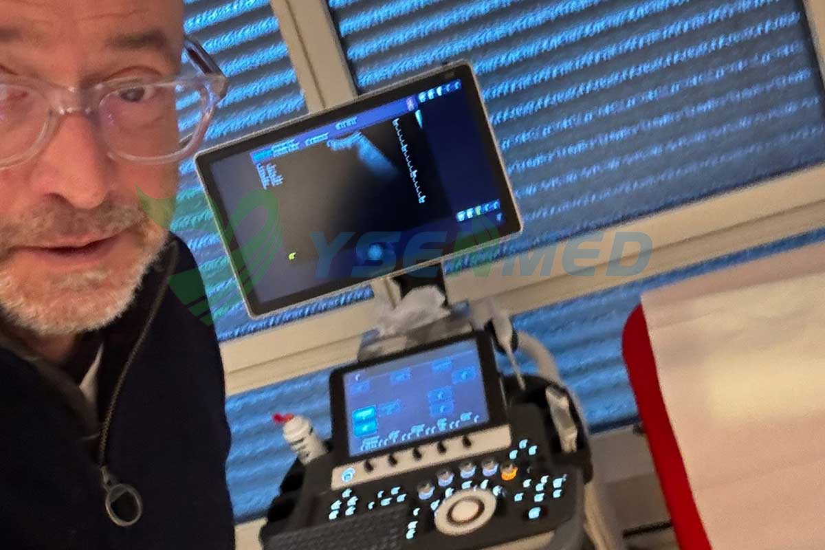
 The well functioning SonoScape S50 ultrasound system makes Dr. Roger happy
Apr .29.2025
Dr. Roger is so excited to receive the SonoScape S50 color doppler ultrasound system, and he can't wait to put it into practice.


