Hot Products
YSX500D 50kW DR system set up and put into service in Cambodia.
YSENMED YSX500D 50kW digital x-ray system has been successfully set up and put into service in a hospital in Cambodia.
YSX056-PE serving as a vehicle-mounted x-ray in the Philippines
YSX056-PE 5.6kW portable x-ray unit has been adapted to fit on a truck, to provide mobile x-ray examination service for remote communities in the Philippines.
X Ray Machine To Zimbabwe
x ray machine, 50KW x ray machine
Microscope To Malawi
Achromatic objectives: 4X、10X、40X(S), 100X(S、Oil) Wide field eyepiece: WF10X(WF16X for option) Eyepiece head: Sliding binocular head inclined at 45° Stage: Double layer mechanical stage size 140X140mm, moving range 75X45mm Focusing: Coaxial coarse and
The Eye's Best Friend: Why Every Clinic Needs an Auto Fundus Camera
Views : 850
Update time : 2025-01-23 17:41:00
When it comes to eye care, precision and technology play pivotal roles. One of the most revolutionary tools in ophthalmology is the auto fundus camera. It's like having a trusty sidekick that never misses a detail. In this article, we'll explore why every clinic should consider adding this incredible device to their arsenal. Let's dive in!

What is an Auto Fundus Camera?
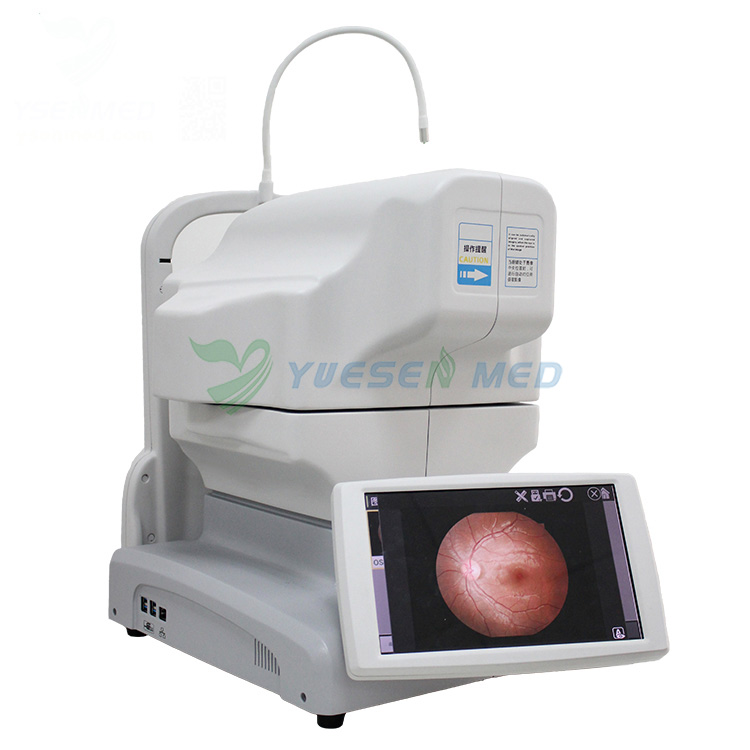
An auto fundus camera is a specialized device designed to capture high-resolution images of the retina. Think of it as a high-tech eye photographer. This camera helps in diagnosing a range of eye conditions by providing clear images of the back of the eye, which includes the retina, optic disc, and macula.
How Does It Work?
The auto fundus camera uses a combination of optics and digital imaging technology. When a patient looks into the camera, it illuminates the eye with a bright light, capturing detailed images without the need for invasive procedures. It's fast, efficient, and, best of all, painless!
The Importance of Retinal Imaging
Early Detection of Eye Diseases
One of the most significant benefits of using an auto fundus camera is its ability to aid in the early detection of eye diseases. Conditions like diabetic retinopathy, glaucoma, and macular degeneration can be detected much earlier with these images. Early detection means early intervention, which can save patients from severe vision loss.
Monitoring Progression of Diseases
For patients already diagnosed with eye conditions, regular imaging allows clinics to monitor the progression of the disease. This ongoing assessment is crucial for adjusting treatment plans and ensuring the best possible outcomes.
Why Clinics Should Invest in an Auto Fundus Camera
Enhanced Patient Care
Investing in an auto fundus camera is like upgrading your clinic's superhero toolkit. It enhances patient care by providing accurate diagnoses and personalized treatment plans. Patients will appreciate the thoroughness and advanced technology, leading to higher satisfaction rates.
Streamlined Workflow
In a busy clinic, time is of the essence. Auto fundus cameras can capture images quickly, allowing for a more streamlined workflow. This efficiency means less waiting time for patients and more time for healthcare providers to focus on what they do best—caring for patients.
Increased Revenue Opportunities
Let's face it: investing in technology can also boost your clinic's bottom line. By offering advanced retinal imaging services, you can attract more patients seeking comprehensive eye care. Additionally, some insurance plans cover these imaging services, creating new revenue streams for your practice.
Features to Look for in an Auto Fundus Camera
Image Quality
When selecting an auto fundus camera, image quality should be a top priority. Look for models that provide high-resolution images, as these are essential for accurate diagnoses.
Ease of Use
The best auto fundus cameras are user-friendly. Clinicians should be able to operate the device with minimal training. Intuitive interfaces and automated features can significantly enhance the user experience.
Portability
Consider the space in your clinic. Some auto fundus cameras are compact and portable, making them easier to integrate into your existing setup without sacrificing valuable floor space.
Connectivity Options
In today's digital age, connectivity is crucial. Look for cameras that offer seamless integration with electronic health record (EHR) systems. This feature allows for easy storage and retrieval of patient images.
The Role of Auto Fundus Cameras in Preventive Care
Educating Patients
Auto fundus cameras not only help in diagnosing conditions but also serve as educational tools. By showing patients images of their eyes, clinicians can explain potential issues and the importance of regular eye check-ups. This visual aid can motivate patients to take their eye health seriously.
Building Trust with Patients
When patients see that their clinic is utilizing advanced technology, it builds trust. They feel confident that they are receiving the best possible care. This trust can lead to long-term relationships and repeat visits.
Overcoming Common Misconceptions
"They're Too Expensive"
While the initial investment in an auto fundus camera may seem daunting, consider the long-term benefits. The cost can be quickly offset by the increased patient volume and enhanced services.
"We Don't Need Advanced Technology"
Some clinics may believe that traditional methods are sufficient. However, in a rapidly evolving field like ophthalmology, staying updated with technology is vital. Patients expect modern solutions, and clinics that adapt will thrive.
Conclusion
In conclusion, an auto fundus camera is more than just a piece of equipment; it's a game-changer for clinics looking to elevate their eye care services. From early disease detection to enhanced patient education, the benefits are undeniable. By investing in this technology, clinics can improve patient outcomes, streamline their workflow, and ultimately grow their practice. So, if you're in the eye care field, ask yourself: can you really afford not to have an auto fundus camera? Your patients—and your practice—will thank you!
FAQ
What is an auto fundus camera used for?
An auto fundus camera is primarily used for capturing detailed images of the retina, optic disc, and macula at the back of the eye. These images help in diagnosing various eye conditions, such as diabetic retinopathy, glaucoma, and macular degeneration. By providing high-resolution images, it enables eye care professionals to assess the health of the retina and monitor any changes over time.
How does an auto fundus camera benefit patients?
The use of an auto fundus camera benefits patients by allowing for early detection of eye diseases, which is crucial for effective treatment. It provides a non-invasive way to examine the retina, making the process quick and painless. Additionally, patients can see images of their own eyes, which helps them understand their conditions better and encourages them to take an active role in their eye health.
Is the procedure with an auto fundus camera painful?
No, the procedure with an auto fundus camera is not painful. Patients simply look into the camera while it captures images of their retina. The bright light used during the imaging may cause temporary discomfort, but there is no physical contact or invasive procedures involved. Most patients find the experience quick and straightforward.
How often should patients have their eyes examined with an auto fundus camera?
The frequency of eye examinations using an auto fundus camera depends on individual risk factors and existing eye conditions. For patients with diabetes or a family history of eye diseases, more frequent examinations may be recommended, typically every 6 to 12 months. For others, a comprehensive eye exam every 1 to 2 years may suffice. It's best to consult with an eye care professional for personalized recommendations.
Related News
Read More >>
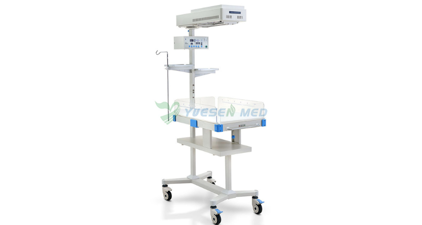
 Why Do Babies Need Infant Radiant Warmers?
Apr .26.2025
One crucial piece of equipment in neonatal care is the infant radiant warmer. But why exactly do babies need these warmers? Let's dive into the world of infant care and explore this important topic.
 Introduction video of YSENMED YSDEN-302S Mobile Dental Chair Unit.
Apr .22.2025
Here we share the introduction video of YSENMED YSDEN-302S Mobile Dental Chair Unit.
 Dr. Mbumba from Gabon highly recommends YSENMED YSX500D DR system
Apr .21.2025
YSENMED has been providing good-valued medical equipment to clinics and hospitals around the world, and we have received a lot of good feedbacks.
DR. Mbumba from Gabon has been advertising our YSX500D digital x-ray system, due to its good performance,
DR. Mbumba from Gabon has been advertising our YSX500D digital x-ray system, due to its good performance,
 What is the Difference Between an Incubator and a Radiant Warmer?
Apr .20.2025
Two essential pieces of equipment often discussed in neonatal care are incubators and radiant warmers. But what exactly sets them apart? Let's dive into the details and explore the reasons.


