


Hot Products
YSX500D 50kW DR system set up and put into service in Cambodia.
YSENMED YSX500D 50kW digital x-ray system has been successfully set up and put into service in a hospital in Cambodia.
YSX056-PE serving as a vehicle-mounted x-ray in the Philippines
YSX056-PE 5.6kW portable x-ray unit has been adapted to fit on a truck, to provide mobile x-ray examination service for remote communities in the Philippines.
X Ray Machine To Zimbabwe
x ray machine, 50KW x ray machine
Microscope To Malawi
Achromatic objectives: 4X、10X、40X(S), 100X(S、Oil) Wide field eyepiece: WF10X(WF16X for option) Eyepiece head: Sliding binocular head inclined at 45° Stage: Double layer mechanical stage size 140X140mm, moving range 75X45mm Focusing: Coaxial coarse and
In Focus: Harnessing the Power of Ultrasonic A/B Scanner for Comprehensive Eye Examinations
Views : 1905
Update time : 2024-05-16 17:01:00
Introduction:
Welcome to the world of eye care where technology continues to push boundaries, providing us with innovative tools to safeguard our most precious sense—sight. In this article, we'll delve into the realm of ultrasonic A/B scanners and explore how they revolutionize the landscape of comprehensive eye examinations. From understanding the basics to unraveling their significance in diagnosing ocular conditions, let's embark on a journey through the lens of cutting-edge ophthalmic technology.
Understanding Ultrasonic A/B Scanners:
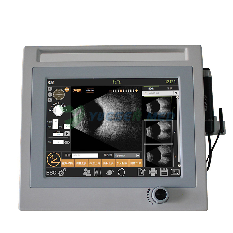
Imagine peering into the intricate layers of the eye with unparalleled precision and clarity. That's precisely what ultrasonic A/B scanners offer. These sophisticated instruments utilize high-frequency sound waves to create detailed images of the eye's internal structures. By emitting pulses of sound and analyzing their echoes, A/B scanners generate real-time images of the anterior and posterior segments of the eye, including the cornea, lens, vitreous, and retina.
The Significance of Comprehensive Eye Examinations:
Before we delve deeper into the mechanics of ultrasonic A/B scanners, let's emphasize the importance of comprehensive eye examinations. Beyond assessing visual acuity, these examinations play a pivotal role in detecting and monitoring various eye conditions, ranging from cataracts and glaucoma to retinal detachment and macular degeneration. Early detection is key to preserving vision and preventing irreversible damage, making regular eye exams a cornerstone of proactive eye care.
Unraveling the Mechanics:
Now, let's unravel the mechanics behind ultrasonic A/B scanners. Unlike traditional imaging techniques like optical coherence tomography (OCT), which rely on light waves, A/B scanners utilize sound waves. The transducer emits ultrasonic pulses directed towards the eye, where they encounter different tissues with varying acoustic properties. These pulses bounce back as echoes, providing valuable insights into the eye's internal structures.
Applications in Ocular Diagnosis:
The versatility of ultrasonic A/B scanners extends to a wide array of ocular diagnoses. From evaluating corneal thickness in refractive surgery candidates to assessing intraocular tumors and foreign bodies, these scanners offer invaluable diagnostic capabilities. Additionally, A/B scans play a crucial role in identifying and characterizing retinal pathologies such as vitreous hemorrhage, retinal detachment, and macular edema, guiding clinicians in formulating tailored treatment plans.
Advantages Over Traditional Imaging Modalities:
While traditional imaging modalities like fundus photography and OCT remain integral to modern ophthalmic practice, ultrasonic A/B scanners offer distinct advantages in certain scenarios. Their ability to penetrate opaque media such as cataracts and vitreous hemorrhages makes them indispensable tools in cases where optical clarity is compromised. Moreover, A/B scans provide comprehensive anatomical information, complementing the qualitative data obtained from other imaging techniques.
Navigating Clinical Challenges:
In the realm of ophthalmology, clinicians often encounter complex clinical scenarios that require meticulous evaluation and precise diagnosis. Ultrasonic A/B scanners prove invaluable in navigating these challenges, offering a non-invasive and reliable means of visualizing ocular structures obscured by media opacities or pathologies. Whether it's differentiating between retinal detachment and vitreous traction or assessing the extent of intraocular tumors, A/B scans empower clinicians with critical insights for informed decision-making.
Enhancing Patient Care:
At the heart of every technological advancement lies a commitment to enhancing patient care. Ultrasonic A/B scanners epitomize this ethos by providing clinicians with the tools they need to deliver personalized and effective treatment strategies. By facilitating early detection, accurate diagnosis, and targeted interventions, these scanners contribute to better outcomes and improved quality of life for patients facing a myriad of ocular conditions.
The Future of Ophthalmic Imaging:
As technology continues to evolve, so too does the landscape of ophthalmic imaging. While ultrasonic A/B scanners have cemented their place as indispensable diagnostic tools, ongoing research and innovation promise even greater advancements in the years to come. From enhanced image resolution to the integration of artificial intelligence algorithms for automated analysis, the future of ophthalmic imaging holds immense promise for optimizing patient care and reshaping the practice of eye care.
Conclusion:
In conclusion, ultrasonic A/B scanners represent a cornerstone of modern ophthalmic practice, offering clinicians unprecedented insights into the intricate anatomy of the eye. From diagnosing ocular pathologies to guiding surgical interventions, these sophisticated instruments empower clinicians with the tools they need to deliver personalized and effective care. As we continue to harness the power of technology in the service of sight, the future of comprehensive eye examinations shines brighter than ever before.
FAQ
How does an ultrasonic A/B scanner differ from other imaging modalities like optical coherence tomography (OCT)?
Ultrasonic A/B scanners utilize sound waves to create images of the eye's internal structures, while OCT relies on light waves. A/B scanners are particularly useful in cases where optical clarity is compromised, as they can penetrate opaque media such as cataracts and vitreous hemorrhages.
What are some common ocular conditions that can be diagnosed using ultrasonic A/B scanners?
Ultrasonic A/B scanners are invaluable in diagnosing a wide range of ocular conditions, including retinal detachment, vitreous hemorrhage, macular edema, intraocular tumors, and foreign bodies. Their versatility and ability to provide comprehensive anatomical information make them indispensable tools in modern ophthalmic practice.
Are ultrasonic A/B scans safe for patients?
Yes, ultrasonic A/B scans are considered safe for patients when performed by trained professionals. The procedure is non-invasive and painless, involving the use of high-frequency sound waves to generate images of the eye's internal structures. However, as with any medical procedure, it's essential to follow appropriate safety protocols and guidelines to minimize risks.
Can ultrasonic A/B scanners be used to monitor the progression of ocular conditions over time?
Yes, ultrasonic A/B scanners play a crucial role in monitoring the progression of ocular conditions by providing detailed images of the eye's internal structures. Clinicians can use these scans to track changes in conditions such as retinal detachment, macular edema, and intraocular tumors, guiding treatment decisions and assessing the efficacy of interventions.
How can ultrasonic A/B scanners contribute to personalized patient care?
Ultrasonic A/B scanners contribute to personalized patient care by providing clinicians with valuable insights into the specific characteristics of ocular conditions. By accurately diagnosing and characterizing these conditions, clinicians can tailor treatment strategies to meet the individual needs of each patient, optimizing outcomes and improving overall quality of care.
Welcome to the world of eye care where technology continues to push boundaries, providing us with innovative tools to safeguard our most precious sense—sight. In this article, we'll delve into the realm of ultrasonic A/B scanners and explore how they revolutionize the landscape of comprehensive eye examinations. From understanding the basics to unraveling their significance in diagnosing ocular conditions, let's embark on a journey through the lens of cutting-edge ophthalmic technology.

Understanding Ultrasonic A/B Scanners:
Imagine peering into the intricate layers of the eye with unparalleled precision and clarity. That's precisely what ultrasonic A/B scanners offer. These sophisticated instruments utilize high-frequency sound waves to create detailed images of the eye's internal structures. By emitting pulses of sound and analyzing their echoes, A/B scanners generate real-time images of the anterior and posterior segments of the eye, including the cornea, lens, vitreous, and retina.
The Significance of Comprehensive Eye Examinations:
Before we delve deeper into the mechanics of ultrasonic A/B scanners, let's emphasize the importance of comprehensive eye examinations. Beyond assessing visual acuity, these examinations play a pivotal role in detecting and monitoring various eye conditions, ranging from cataracts and glaucoma to retinal detachment and macular degeneration. Early detection is key to preserving vision and preventing irreversible damage, making regular eye exams a cornerstone of proactive eye care.
Unraveling the Mechanics:
Now, let's unravel the mechanics behind ultrasonic A/B scanners. Unlike traditional imaging techniques like optical coherence tomography (OCT), which rely on light waves, A/B scanners utilize sound waves. The transducer emits ultrasonic pulses directed towards the eye, where they encounter different tissues with varying acoustic properties. These pulses bounce back as echoes, providing valuable insights into the eye's internal structures.
Applications in Ocular Diagnosis:
The versatility of ultrasonic A/B scanners extends to a wide array of ocular diagnoses. From evaluating corneal thickness in refractive surgery candidates to assessing intraocular tumors and foreign bodies, these scanners offer invaluable diagnostic capabilities. Additionally, A/B scans play a crucial role in identifying and characterizing retinal pathologies such as vitreous hemorrhage, retinal detachment, and macular edema, guiding clinicians in formulating tailored treatment plans.
Advantages Over Traditional Imaging Modalities:
While traditional imaging modalities like fundus photography and OCT remain integral to modern ophthalmic practice, ultrasonic A/B scanners offer distinct advantages in certain scenarios. Their ability to penetrate opaque media such as cataracts and vitreous hemorrhages makes them indispensable tools in cases where optical clarity is compromised. Moreover, A/B scans provide comprehensive anatomical information, complementing the qualitative data obtained from other imaging techniques.
Navigating Clinical Challenges:
In the realm of ophthalmology, clinicians often encounter complex clinical scenarios that require meticulous evaluation and precise diagnosis. Ultrasonic A/B scanners prove invaluable in navigating these challenges, offering a non-invasive and reliable means of visualizing ocular structures obscured by media opacities or pathologies. Whether it's differentiating between retinal detachment and vitreous traction or assessing the extent of intraocular tumors, A/B scans empower clinicians with critical insights for informed decision-making.
Enhancing Patient Care:
At the heart of every technological advancement lies a commitment to enhancing patient care. Ultrasonic A/B scanners epitomize this ethos by providing clinicians with the tools they need to deliver personalized and effective treatment strategies. By facilitating early detection, accurate diagnosis, and targeted interventions, these scanners contribute to better outcomes and improved quality of life for patients facing a myriad of ocular conditions.
The Future of Ophthalmic Imaging:
As technology continues to evolve, so too does the landscape of ophthalmic imaging. While ultrasonic A/B scanners have cemented their place as indispensable diagnostic tools, ongoing research and innovation promise even greater advancements in the years to come. From enhanced image resolution to the integration of artificial intelligence algorithms for automated analysis, the future of ophthalmic imaging holds immense promise for optimizing patient care and reshaping the practice of eye care.
Conclusion:
In conclusion, ultrasonic A/B scanners represent a cornerstone of modern ophthalmic practice, offering clinicians unprecedented insights into the intricate anatomy of the eye. From diagnosing ocular pathologies to guiding surgical interventions, these sophisticated instruments empower clinicians with the tools they need to deliver personalized and effective care. As we continue to harness the power of technology in the service of sight, the future of comprehensive eye examinations shines brighter than ever before.
FAQ
How does an ultrasonic A/B scanner differ from other imaging modalities like optical coherence tomography (OCT)?
Ultrasonic A/B scanners utilize sound waves to create images of the eye's internal structures, while OCT relies on light waves. A/B scanners are particularly useful in cases where optical clarity is compromised, as they can penetrate opaque media such as cataracts and vitreous hemorrhages.
What are some common ocular conditions that can be diagnosed using ultrasonic A/B scanners?
Ultrasonic A/B scanners are invaluable in diagnosing a wide range of ocular conditions, including retinal detachment, vitreous hemorrhage, macular edema, intraocular tumors, and foreign bodies. Their versatility and ability to provide comprehensive anatomical information make them indispensable tools in modern ophthalmic practice.
Are ultrasonic A/B scans safe for patients?
Yes, ultrasonic A/B scans are considered safe for patients when performed by trained professionals. The procedure is non-invasive and painless, involving the use of high-frequency sound waves to generate images of the eye's internal structures. However, as with any medical procedure, it's essential to follow appropriate safety protocols and guidelines to minimize risks.
Can ultrasonic A/B scanners be used to monitor the progression of ocular conditions over time?
Yes, ultrasonic A/B scanners play a crucial role in monitoring the progression of ocular conditions by providing detailed images of the eye's internal structures. Clinicians can use these scans to track changes in conditions such as retinal detachment, macular edema, and intraocular tumors, guiding treatment decisions and assessing the efficacy of interventions.
How can ultrasonic A/B scanners contribute to personalized patient care?
Ultrasonic A/B scanners contribute to personalized patient care by providing clinicians with valuable insights into the specific characteristics of ocular conditions. By accurately diagnosing and characterizing these conditions, clinicians can tailor treatment strategies to meet the individual needs of each patient, optimizing outcomes and improving overall quality of care.
Related News
Read More >>
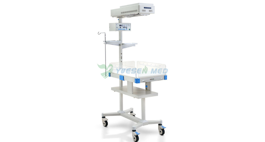 Why Do Babies Need Infant Radiant Warmers?
Apr .26.2025
One crucial piece of equipment in neonatal care is the infant radiant warmer. But why exactly do babies need these warmers? Let's dive into the world of infant care and explore this important topic.
 Introduction video of YSENMED YSDEN-302S Mobile Dental Chair Unit.
Apr .22.2025
Here we share the introduction video of YSENMED YSDEN-302S Mobile Dental Chair Unit.
 Dr. Mbumba from Gabon highly recommends YSENMED YSX500D DR system
Apr .21.2025
YSENMED has been providing good-valued medical equipment to clinics and hospitals around the world, and we have received a lot of good feedbacks.
DR. Mbumba from Gabon has been advertising our YSX500D digital x-ray system, due to its good performance,
DR. Mbumba from Gabon has been advertising our YSX500D digital x-ray system, due to its good performance,
 What is the Difference Between an Incubator and a Radiant Warmer?
Apr .20.2025
Two essential pieces of equipment often discussed in neonatal care are incubators and radiant warmers. But what exactly sets them apart? Let's dive into the details and explore the reasons.
