


Hot Products
YSX500D 50kW DR system set up and put into service in Cambodia.
YSENMED YSX500D 50kW digital x-ray system has been successfully set up and put into service in a hospital in Cambodia.
YSX056-PE serving as a vehicle-mounted x-ray in the Philippines
YSX056-PE 5.6kW portable x-ray unit has been adapted to fit on a truck, to provide mobile x-ray examination service for remote communities in the Philippines.
X Ray Machine To Zimbabwe
x ray machine, 50KW x ray machine
Microscope To Malawi
Achromatic objectives: 4X、10X、40X(S), 100X(S、Oil) Wide field eyepiece: WF10X(WF16X for option) Eyepiece head: Sliding binocular head inclined at 45° Stage: Double layer mechanical stage size 140X140mm, moving range 75X45mm Focusing: Coaxial coarse and
Through the Lens: Understanding the Role of Auto Fundus Camera in Ophthalmology
Views : 1887
Update time : 2024-05-17 15:42:00
In the vast world of ophthalmology, where the intricate complexities of the human eye meet cutting-edge technology, the auto fundus camera stands out as a remarkable tool. Its significance lies not only in its ability to capture high-resolution images of the fundus but also in its potential to revolutionize diagnosis and treatment protocols. Let's delve into the fascinating world of auto fundus cameras and explore their indispensable role in modern ophthalmic practice.
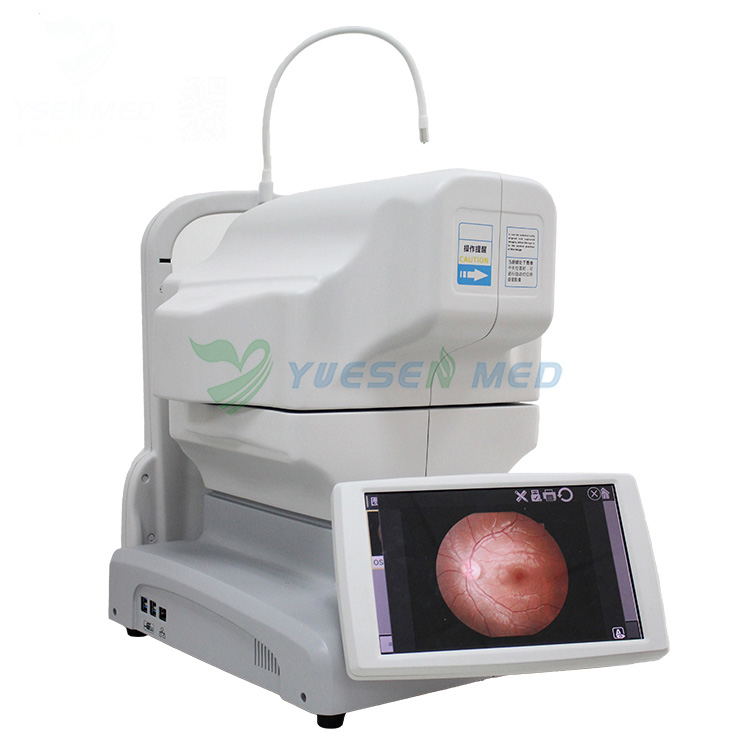
What is an Auto Fundus Camera?
At its core, an auto fundus camera is a specialized imaging device designed to capture detailed images of the fundus—the interior surface of the eye, including the retina, optic disc, and blood vessels. Unlike traditional fundus cameras that require manual adjustments and expertise, auto fundus cameras employ advanced technology to automate the imaging process, making it faster, more efficient, and less reliant on operator skill.
The Technology Behind Auto Fundus Cameras
Auto fundus cameras utilize a combination of sophisticated optics, digital sensors, and intelligent software algorithms to achieve precise imaging results. These cameras feature high-resolution sensors capable of capturing detailed images with exceptional clarity and contrast. Advanced autofocus mechanisms ensure optimal focus on the target structures, eliminating the need for manual adjustments and reducing the risk of image blurring or distortion.
Applications in Ophthalmology
The applications of auto fundus cameras in ophthalmology are vast and varied, ranging from routine screening and diagnosis to monitoring disease progression and guiding treatment decisions. Here are some key areas where auto fundus cameras play a crucial role:
Diabetic Retinopathy Screening
Diabetic retinopathy is a common complication of diabetes and a leading cause of blindness worldwide. Early detection and timely intervention are essential for preventing vision loss in affected individuals. Auto fundus cameras facilitate efficient diabetic retinopathy screening programs by enabling rapid imaging of the retina and early identification of characteristic changes such as microaneurysms, hemorrhages, and exudates.
Glaucoma Management
Glaucoma is a progressive optic neuropathy characterized by damage to the optic nerve and visual field loss. Monitoring changes in the optic disc and retinal nerve fiber layer is crucial for assessing disease progression and evaluating the effectiveness of treatment interventions. Auto fundus cameras provide detailed imaging of the optic disc and retinal structures, allowing ophthalmologists to track subtle changes over time and adjust treatment strategies accordingly.
Age-Related Macular Degeneration (AMD)
AMD is a leading cause of irreversible vision loss in older adults, affecting the macula—the central part of the retina responsible for sharp, central vision. Auto fundus cameras play a vital role in diagnosing and monitoring AMD by capturing detailed images of macular changes such as drusen, pigmentary alterations, and geographic atrophy. Early detection of AMD allows for timely intervention with treatments such as anti-VEGF therapy or photodynamic therapy, aimed at preserving vision and preventing disease progression.
Retinopathy of Prematurity (ROP)
ROP is a potentially blinding disease that affects premature infants, characterized by abnormal blood vessel growth in the retina. Screening for ROP is essential to identify infants at risk of vision loss and initiate timely interventions such as laser therapy or anti-VEGF injections. Auto fundus cameras enable non-invasive imaging of the immature retina, facilitating early detection and appropriate management of ROP in neonatal intensive care units.
Advantages of Auto Fundus Cameras
The adoption of auto fundus cameras in ophthalmic practice offers several advantages over traditional imaging methods:
- Efficiency: Auto fundus cameras streamline the imaging process, reducing the time and resources required for capturing high-quality images.
- Consistency: Automated imaging protocols ensure consistency in image acquisition, minimizing variability and enhancing reliability for diagnostic interpretation.
- Accessibility: Auto fundus cameras can be deployed in various clinical settings, including primary care offices, community health centers, and remote locations, expanding access to retinal screening services for underserved populations.
- Patient Comfort: The non-invasive nature of auto fundus imaging enhances patient comfort and compliance, particularly in pediatric and elderly populations.
Future Perspectives
As technology continues to evolve, the future of auto fundus cameras holds exciting possibilities for further advancements in imaging quality, automation capabilities, and diagnostic algorithms. Integration with artificial intelligence (AI) algorithms for image analysis and interpretation promises to enhance diagnostic accuracy and efficiency, paving the way for personalized medicine approaches in ophthalmology.
Conclusion
In conclusion, auto fundus cameras represent a cornerstone of modern ophthalmic practice, offering unparalleled capabilities for retinal imaging and disease management. From diabetic retinopathy screening to glaucoma management and beyond, these innovative devices empower clinicians with valuable insights into ocular health and facilitate early intervention to preserve vision and improve patient outcomes. As we look ahead, the continued advancement of auto fundus camera technology holds immense promise for shaping the future of eye care and transforming the landscape of ophthalmology.
FAQs:
How does an auto fundus camera differ from a traditional fundus camera?
Auto fundus cameras utilize advanced technology to automate the imaging process, eliminating the need for manual adjustments and operator expertise. Traditional fundus cameras require skilled operators to adjust settings such as focus, aperture, and illumination, whereas auto fundus cameras employ intelligent algorithms to achieve optimal imaging results automatically. This automation enhances efficiency, reduces variability, and improves consistency in image acquisition, making it an ideal choice for routine screening and diagnosis in ophthalmic practice.
Are auto fundus cameras suitable for pediatric patients?
Yes, auto fundus cameras are well-suited for imaging pediatric patients, offering non-invasive and patient-friendly imaging solutions. The automated nature of these cameras minimizes the need for prolonged image acquisition, reducing the likelihood of patient discomfort or agitation. Additionally, the high-resolution imaging capabilities of auto fundus cameras enable detailed visualization of the pediatric retina, facilitating the early detection and management of conditions such as retinopathy of prematurity (ROP) and congenital retinal disorders.
How do auto fundus cameras contribute to diabetic retinopathy screening?
Auto fundus cameras play a vital role in diabetic retinopathy screening programs by enabling efficient and accurate imaging of the retina. These cameras facilitate the early detection of characteristic changes such as microaneurysms, hemorrhages, and exudates, allowing for timely intervention to prevent vision loss in diabetic patients. The automated nature of auto fundus cameras streamlines the screening process, making it more accessible and cost-effective for healthcare providers and patients alike.
Can auto fundus cameras assist in the management of glaucoma?
Yes, auto fundus cameras are valuable tools for the management of glaucoma, providing detailed imaging of the optic disc and retinal nerve fiber layer. By monitoring changes in these structures over time, ophthalmologists can assess disease progression, evaluate treatment effectiveness, and make informed decisions regarding patient care. The automated imaging protocols of auto fundus cameras ensure consistency and reliability in image acquisition, enhancing diagnostic accuracy and facilitating personalized treatment strategies for glaucoma patients.
What are the advantages of using auto fundus cameras in ophthalmic practice?
The adoption of auto fundus cameras offers several advantages for ophthalmic practice, including efficiency, consistency, accessibility, and patient comfort. These cameras streamline the imaging process, reducing the time and resources required for capturing high-quality images. Automated imaging protocols ensure consistency in image acquisition, minimizing variability and enhancing reliability for diagnostic interpretation. Auto fundus cameras can be deployed in various clinical settings, expanding access to retinal screening services for underserved populations. Additionally, the non-invasive nature of auto fundus imaging enhances patient comfort and compliance, particularly in pediatric and elderly populations.
Related News
Read More >>
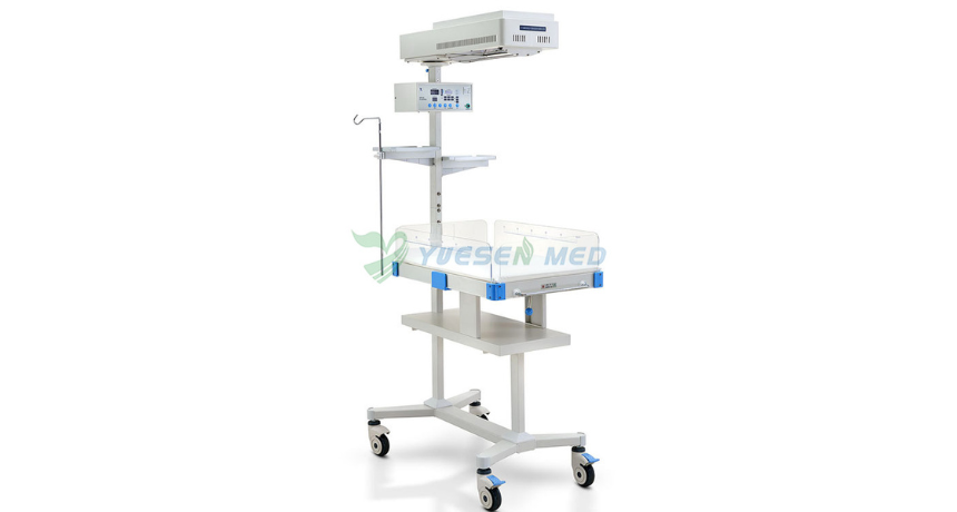 Why Do Babies Need Infant Radiant Warmers?
Apr .26.2025
One crucial piece of equipment in neonatal care is the infant radiant warmer. But why exactly do babies need these warmers? Let's dive into the world of infant care and explore this important topic.
 Introduction video of YSENMED YSDEN-302S Mobile Dental Chair Unit.
Apr .22.2025
Here we share the introduction video of YSENMED YSDEN-302S Mobile Dental Chair Unit.
 Dr. Mbumba from Gabon highly recommends YSENMED YSX500D DR system
Apr .21.2025
YSENMED has been providing good-valued medical equipment to clinics and hospitals around the world, and we have received a lot of good feedbacks.
DR. Mbumba from Gabon has been advertising our YSX500D digital x-ray system, due to its good performance,
DR. Mbumba from Gabon has been advertising our YSX500D digital x-ray system, due to its good performance,
 What is the Difference Between an Incubator and a Radiant Warmer?
Apr .20.2025
Two essential pieces of equipment often discussed in neonatal care are incubators and radiant warmers. But what exactly sets them apart? Let's dive into the details and explore the reasons.
